To provide the highest standard of care, our practice is equipped with state-of-the-art technology designed to streamline and accelerate both diagnosis and treatment.
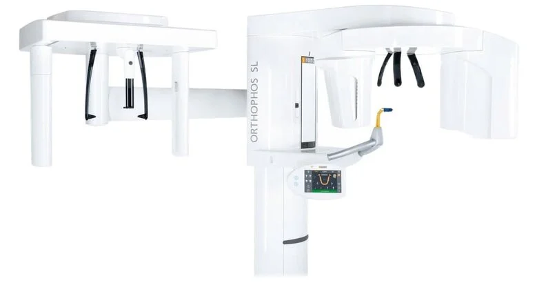
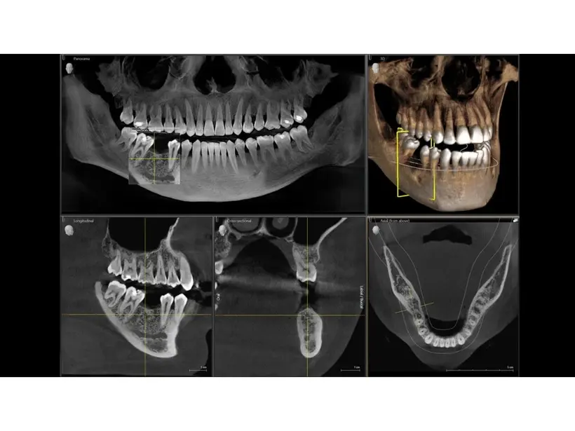
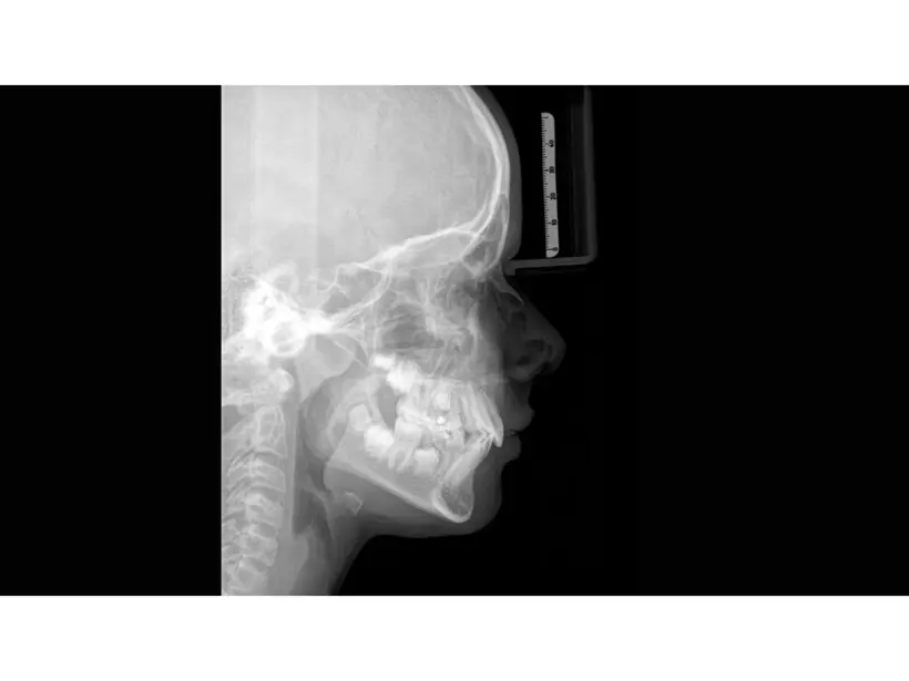
Imaging Diagnostics: 2D and 3D Radiology
In our office, we can perform intraoral X-rays, panoramic radiographs, lateral cephalometric X-rays, and CBCT scans using a single advanced system that integrates all of these functions.
These exams help us detect cavities, evaluate bone health, identify problems that may not be visible during a clinical exam, confirm the presence or absence of teeth, and plan surgical procedures and orthodontic treatment with greater precision.
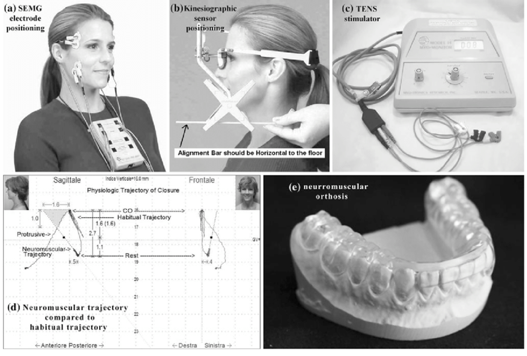
Cranio-Mandibular Kinesiography
Cranio-mandibular kinesiography is one of our most frequently used diagnostic tools for patients with TMJ (temporomandibular joint) concerns—and not only for TMJ. By combining kinesiography, surface electromyography (sEMG), and ULF-TENS (ultra–low-frequency TENS), specialized software generates detailed graphs that map how the jaw system is functioning. This allows our doctors to reach a diagnosis with sub-millimeter precision, down to tenths of a millimeter.
It is the only tool that provides a truly dynamic diagnosis: instead of relying solely on static measurements, it records how the patient functions in real time, showing what happens during jaw movements—and therefore what needs to be corrected to restore healthy function.
Kinesiography is rooted in the work of Dr. Bernard Jankelson, considered the founder of neuromuscular dentistry. He famously stated that “whatever can be measured is fact; everything else is opinion”—a principle we strongly value.
The exam takes approximately two hours and can be performed for both adults and children.
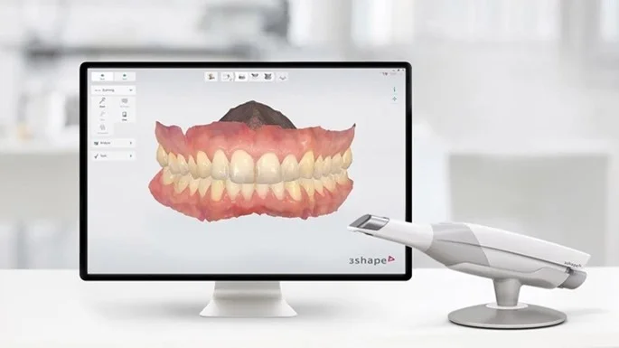
3D Impressions
Remember how uncomfortable traditional alginate impressions could be? You can forget about that.
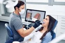
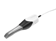
Intraoral scanning is a true game-changer in dentistry. Using a digital scanner, we capture a series of high-definition images of your mouth, which are then processed by specialized software to create a realistic, highly accurate 3D model of your teeth.
The process is completely painless, fast, and exceptionally precise.
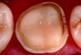
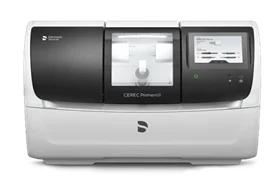
The CEREC CAD/CAM System
CAD/CAM technology (Computer-Aided Design and Computer-Aided Manufacturing) is used when a tooth has lost a significant amount of structure due to decay, wear, fracture, or previous dental work. With this system, we can digitally design and manufacture highly precise restorations in minutes—recreating the missing portion of one or more teeth with exceptional accuracy.
CEREC was originally developed for inlays and crowns, but it can also be used to produce veneers, as well as temporary and definitive restorations. In many cases, everything can be completed in a single appointment, eliminating traditional lab wait times.
Achieving the final result typically involves five steps:
1. Tooth preparation: the dentist prepares the surface of the tooth to be restored, following precise clinical parameters.
2. Optical scanning: using a scanner integrated into the system, we capture 3D digital impressions of the mouth, with particular focus on the tooth that needs to be restored.
3. Design: the CEREC software is fast and highly intuitive. We digitally design the restoration for the prepared tooth, customizing it to the patient’s unique anatomy.
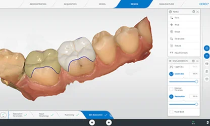
4. Milling (Production): the unit receives the design from the software and, using precision burs, “sculpts” the restoration from a ceramic or composite block in the selected shade—delivering strength and aesthetics in as little as 15 minutes. It’s essentially the practice’s robotic Michelangelo.
5. Bonding (Cementation): the restoration is bonded to the tooth that was prepared at the start. If needed, it can be refined directly in the mouth to ensure the best possible fit and bite.
Nitrous Oxide Sedation (Conscious Sedation) System
Henry Langa is credited with developing this technique in the 1970s, and it quickly became widely adopted in English-speaking countries. This equipment allows us to administer a carefully controlled mixture of nitrous oxide (the well-known “laughing gas”) and oxygen in complete safety, helping patients reach a state of conscious sedation with relative analgesia—particularly useful for reducing dental anxiety and fear.
Dental phobia affects an estimated 3.5–5% of patients, many of whom avoid necessary treatment because their anxiety feels impossible to overcome.
It’s important not to confuse this technique with general anesthesia: the patient remains awake and responsive, continuously monitored (including oxygen saturation), but experiences a pleasant sense of calm that makes it easier to stay relaxed and cooperative during care. There is nothing to be embarrassed about—this approach helps both the patient and the clinician establish a more comfortable, positive routine of treatment.